BxLink™ (exclusive to Lumea):
BxLink is an all-in-one solution that offers everything from billing to preliminary reporting for practices ready to go digital without changing their existing LIS. It streamlines workflows with biopsy tracking, paperless lab requisitions, and seamless pathology work lists, backed by RFID specimen tracking for added security. BxLink tracks tissue at every step of the diagnostic process, providing real-time updates, such as when the lab receives a sample. Labs can integrate it with or use it as a replacement for their LIS. Then, pathologists can use it to view digital slides, annotate findings, and generate final reports, which are instantly communicated to the clinic.
With easy integration into any EMR and advanced AI algorithms, the platform simplifies report sharing and enhances research opportunities. It also supports digital test orders, faster result integration, and efficient sample tracking, improving both clinic and lab workflows.

BxBoard® (exclusive to Lumea):
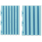
The BxBoard uses patented tissue preservation technology to safely transport samples from clinic to lab, replacing the outdated method of free-floating samples in formalin bottles, which could lead to issues like curling or over-fixation. Each BxBoard features either six or four lanes to securely hold core needle biopsies, preserving tissue orientation and reducing the risk of segmentation. This compact device consolidates what would typically require multiple bottles into one, reducing the risk of specimen mix-ups and reducing formaldehyde exposure for medical staff.
BxLink tracks the BxBoard via RFID tags, notifying labs when samples are in transit and automatically pulling up patient information when scanned in the lab, keeping processes streamlined and organized.

BxChip® (exclusive to Lumea):
Once BxBoards arrive at a lab, the cores can be transferred from the board to the BxChip. The BxChip is a patented sectional matrix designed to hold four or six core biopsies during lab processing, keeping tissues flat on an even plane and maintaining orientation for optimal review. Its design improves tissue orientation and integrity and, in some labs, has resulted in a 14.5% increase in tissue surface area, a 31.8% increase in core length, and a 9.3% increase in cancer detection rates.

BxCamera® (exclusive to Lumea):
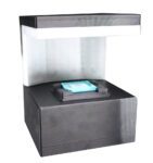
Once prostate or other specimens have been transferred from the BxBoard to the BxChip, they can be easily and quickly grossed with the BxCamera. The BxCamera is an AI-powered tool designed to assist in grossing by quickly measuring and documenting tissue specimens. Lab staff simply have to place the chip under the camera, and the system uses machine learning to measure the surface area of the tissue in seconds, improving both efficiency and accuracy in specimen processing.

Digital Slide Scanner:
In the last step of the digital pathology prostate tissue process in the lab, a digital slide scanner will upload the samples to BxLink. Pathologists can view, annotate, and diagnose digitally from any validated location, which is very helpful in a world where remote work has become common.

Example: Leica Digital Slide Scanner
Artificial Intelligence/AI:
Artificial intelligence is used in digital pathology to analyze the tissue cores uploaded to the digital software, or BxLink. After making a diagnosis, a pathologist can use the AI integrated with BxLink to review their diagnosis or patterns too small for the human eye to detect.

All of this technology combined has significantly improved cancer detection, workflows, and efficiency for many groups worldwide. To learn more about Lumea’s technology for prostate and other types of cancer, request more information from us today.

