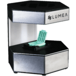
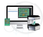
What is the BxCamera?
The BxCamera is an AI-assisted grossing tool for simple biopsy imaging and measuring. This camera works with Lumea’s tissue transportation devices in the surgery center or the lab. It uses machine learning to localize, count, and measure each tissue fragment and provide a gross description that can auto-populate into a template within the LIS.

What are the benefits of using the BxCamera?
- Labs have found that using the BxCamera saves over 62% of grossing time.
- BxCamera’s advanced camera algorithms have been used to image more than 2 million specimens.
- The camera can detect, measure, and provide a gross description of anything that fits in a cassette, including multiple pieces of tissue.
- Machine learning auto-measures and documents tissue that populates into a template or MACRO, saving valuable time.
- When used at the procedure site, it becomes an asset in the chain of custody tracking. Labs can use the image taken at the procedure site as extra verification.
- The microtomist can compare the block appearance to the grossing image as a quality assurance step to verify the full face of the specimen.
How does the BxCamera help labs standardize their histology processes?
When a lab implements the BxCamera in their grossing process, they can gain significant improvements in standardized tissue handling. Instead of using a ruler to meticulously measure specimens (which introduces variability), approved lab staff can capture an image of the tissue and instantly populate its size into a report or LIS.
The BxCamera also makes it easy to provide grossing information in the same way each time a specimen is analyzed, streamlining all subsequent processes and outputs.
Where can the BxCamera be used?
The camera can be used at the procedure site or in the laboratory. When used at the procedure, lab personnel validate the image when they receive the specimen.
Which specimen sizes can the BxCamera image?
The BxCamera was created for small tissue specimens. Our Research and Development department is currently working on a version for larger specimens.
Do I need to use Lumea’s software with the BxCamera?
No—the BxCamera can be used as a standalone solution without any LIS integrations.
If you’re interested in integrating the BxCamera into your current LIS (allowing grossing staff to use the camera and transfer completed gross statements automatically back into the existing LIS report) or adopting Lumea software, you can contact us here for more information.
What’s included in a BxCamera camera kit?
- Camera
- Power Supply Head
- USB-C to C Cable
- USB-C to Camera Cord
- Ethernet Cord
- USB to Ethernet Adapter
- Cassette Platform
- BxView Platform
- Cassette Adapter
- Calibration Target with Replaceable Calibration Papers
Is it FDA-registered?
Yes! The BxCamera is registered with the FDA.

A 12-core prostate biopsy case can take anywhere between 30 minutes to an hour. Using Lumea technology, the same case can be accessioned and grossed in five to ten minutes. “We used to have to dictate into a microphone the numbers as we hand-measured each specimen. Now, we put the specimen into this little camera and it does all the work for us,” said Ann Mazurco, a histotechnologist.
Learn more about what the BxCamera can do for your practice by reading our article on how it can improve accessioning and grossing.


