Welcome to our FAQ blog post about the Lumea BxChip. Here, we will answer some of the common questions you may have about this helpful technology for labs and pathologists.
 1. Product Overview & Compatibility
What is the BxChip?
The BxChip is a patented, engineered, tissue-mimetic array designed to safely hold and manage multiple core needle biopsies in a single cassette throughout tissue processing. It standardizes tissue handling and reduces cassette and block volume in the lab.
What is the BxChip made for?
The BxChip is made for small core needle biopsies.
What are the available BxChip options?
Lumea offers several options tailored to different biopsy types and sizes:
- BxChip – 6-Lane: Compatible with 16–18 gauge core needle biopsies (ideal for prostate core biopsies).
- BxChip – 4-Lane: Compatible with 11-14 gauge core needle biopsies (ideal for larger gauge breast core biopsies).
- In Development: BxChip – 8 Well (up to 7mm tissue) and BxChip – 16 Well (up to 4mm tissue).
2. Clinical & Operational Benefits
The BxChip provides significant improvements in both tissue quality and lab efficiency.
Clinical Impact
- Increased Tissue Yield: The BxChip reduces linear variability, resulting in an average 31.8% increase in biopsy core length and a 14.5% average increase in histologic tissue surface area on the glass slide.
- Enhanced Detection: Pathologists using the BxChip have reported an average of up to an 18.79% increase in overall cancer detection rate.
- Cost Savings in Staining: Multiplexing up to six cores on one slide enables labs to perform additional stains with significantly reduced costs.

Operational Efficiency
The multiplexing and standardization provided by the BxChip lead to substantial time savings for grossing and embedding, as well as other savings on materials, cost, and storage space because of reduced cassette and block volume.
3. Lab Workflow & Functionality
How does the BxChip work during tissue processing?
The BxChip material mimics the properties of human tissue, allowing it to process, embed, cut, and stain seamlessly. During processing, the BxChip is formulated to shrink down slightly to maintain the core specimens in their fixed position. Once stained, the material will appear as a background on the slide, maintaining the original placement of the tissues.
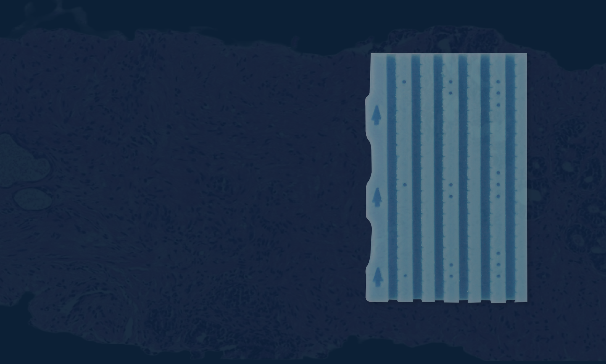
How is core orientation and identity maintained?
BxChips contain special markings to guide the pathologist:
- Orientation: Arrows indicate the proximal and distal ends of the tissue.
- Identification: Dots serve as a key to identify which core was placed where (from left to right).
Labs should document which tissue corresponds to which designation. If used with Lumea’s software, our technology will automatically track and maintain these designations.
How does the BxChip improve slide scanning?
The distinct blue material of the BxChip acts as an anchor, allowing digital scanners to lock in and focus on the relevant tissue areas, improving scanned slide quality.
How do pathologists diagnose with the BxChip?
Pathologists diagnose the cores within the BxChip by using the core identification key. If used with Lumea’s Viewer+™ platform, pathologists can view and annotate cores digitally, easily toggle between different stains, and our tissue-detection software will auto-measure tissue involvement and draft a diagnostic report.
4. Logistics & Integration
How does the BxChip integrate with other Lumea technology?
The BxChip works effectively as a standalone product but is optimized for the Lumea ecosystem:
- BxCamera®: Quickly and accurately captures an image and measures tissue in the BxChip during grossing.
- BxBoard®: Used by procedure facilities instead of formalin jars. It helps maintain specimen orientation and keeps cores flat and straight, facilitating an ideal transfer to the BxChip.
- Viewer+™ & BxLink™ Platforms: Our software uses the chip as a landmark for AI, allowing multiple slides and levels of the same core to be viewed simultaneously on an aligned grid.
Is the BxChip regulatory compliant?
Yes. The BxChip is registered with the FDA and holds the CE marking, which certifies compliance with EU health, safety, and environmental requirements.

What are you waiting for? Buy our BxChip onboarding kit today to try it in your lab. Want to learn more? Request more information from our team.